Associação Portuguesa de Investigação em Cancro
Obesity promotes the progression of prostate cancer
Obesity promotes the progression of prostate cancer

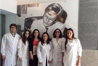
A researcher group of the Unit for Multidisciplinary Research in Biomedicine (UMIB) led by Professor Mariana P. Monteiro developed a research project, co-financed by the Portuguese Society of Endocrinology Diabetes and Metabolism (SPEDM), whose objective was to evaluate the influence of obesity in androgen independent prostate carcinoma progression. For this purpose, in vitro studies were performed in order to evaluate the effect of factors released by adipocytes and preadipocytes in proliferation, migration and invasion of androgen independent prostate carcinoma cells. It was observed that the factors released by adipocytes lead to an increase in the viability, proliferation, migration and invasion of prostate carcinoma cells, suggesting that obesity could promote the progression and aggressiveness of this tumor.
Authors and Affiliations:
Ângela Moreira1, MSc; Sofia S Pereira1, MSc; Madalena Costa1, BSc; Tiago Morais1, MSc; Ana Pinto1, Bsc; Ruben Fernandes2, PhD; Mariana P. Monteiro1, MD, PhD.
1 Department of Anatomy, Unit for Multidisciplinary Research in Biomedicine (UMIB), Institute for Biomedical Sciences Abel Salazar (ICBAS), University of Porto, 4050-313 Porto , Portugal.
2 Ciências Químicas e das Biomoléculas (CQB) e Centro de Investigação em Saúde e Ambiente (CISA), Escola Superior de Tecnologia da Saúde do Porto do Instituto Politécnico do Porto (ESTSP-IPP), 4400-330 Vila Nova de Gaia, Portugal.
Abstract:
Obesity has been associated with increased incidence and risk of mortality of prostate cancer. One of the proposed mechanisms underlying this risk association is the changes in adipokines expression that could promote the development and progression of the prostate tumor cells.
The main goal of this study was to evaluate the effect of preadipocyte and adipocyte secretome in the proliferation, migration and invasion of androgen independent prostate carcinoma cells (RM1) and to assess cell proliferation in the presence of the adiposity signals leptin and insulin.
RM1 cells were co-cultured in with preadipocytes, adipocytes or cultured in their respective conditioned medium. Cell proliferation was assessed by flow cytometry and XTT viability test. Cell migration was evaluated using a wound healing injury assay of RM1 cells cultured with conditioned media. Cellular invasion of RM1 cells co-cultured with adipocytes and preadipocytes was assessed using matrigel membranes.
Preadipocyte conditioned medium were associated with a small increase in RM1 proliferation, while adipocytes conditioned media significantly increased RM1 cell proliferation (p<0.01). Adipocytes also significantly increased the RM1 cells proliferation in co-culture (p <0.01). Cell migration was higher in RM1 cells cultured with preadipocyte and adipocyte conditioned medium. RM1 cell invasion was significantly increased after co-culture with preadipocytes and adipocytes (p <0.05). Insulin also increased significantly the cell proliferation in contrast to leptin, which showed no effect.
In conclusion, prostate carcinoma cells seem to be influenced by factors secreted by adipocytes that are able to increase their ability to proliferate, migrate and invade.
Journal: PLOS ONE
Link:http://journals.plos.org/plosone/article?id=10.1371/journal.pone.0123217


