Associação Portuguesa de Investigação em Cancro
MEK5-ERK5 signaling contributes to the progression of colon cancer
MEK5-ERK5 signaling contributes to the progression of colon cancer
AES Simões1, DM Pereira1, SE Gomes1, H Brito1, T Carvalho2, A French3, RE Castro1, CJ Steer4,5, SN Thibodeau3, CMP Rodrigues*,1,6 and PM Borralho*,1,6
1 Research Institute for Medicines (iMed.ULisboa), Faculty of Pharmacy, Universidade de Lisboa, Av. Prof. Gama Pinto, Lisbon 1649-003, Portugal;
2 Histology and Comparative Pathology Laboratory, Instituto de Medicina Molecular, Av. Prof. Egas Moniz, Edificio Egas Moniz, Lisbon 1649-028, Portugal;
3 Department of Laboratory Medicine and Pathology, Mayo Clinic, 200 First Street S.W., Rochester, MN 55905, USA;
4 Department of Medicine, VFW Cancer Research Center, University of Minnesota Medical School, 406 Harvard Street S.E., Minneapolis, MN 55455, USA
5 Department of Genetics, Cell Biology and Development, VFW Cancer Research Center, University of Minnesota Medical School, 406 Harvard Street S.E., Minneapolis, MN 55455, USA
* Corresponding authors: CMP Rodrigues or PM Borralho, Research Institute for Medicines (iMed.ULisboa), Faculty of Pharmacy, Universidade de Lisboa, Av. Prof. Gama Pinto, Lisbon 1649-003, Portugal. Tel: +351 217 946 400; Fax: +351 21 794 6491; E-mail: [email protected] or [email protected]
6 These authors are the co-senior authors.
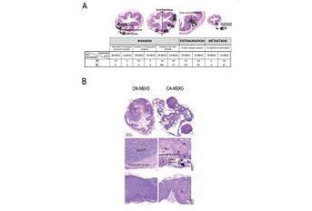
This study was designed to evaluate MEK5 and ERK5 expression in colon cancer progression and to ascertain the relevance of MEK5/ERK5 signalling in colon cancer. Expression of MEK5 and ERK5 was evaluated in 323 human colon cancer samples. To evaluate the role of MEK5/ERK5 signalling in colon cancer, we developed a stable cell line model with differential MEK5/ERK5 activation. Impact of differential MEK5/ERK5 signalling was evaluated on cell cycle progression by flow cytometry and cell migration was evaluated by wound healing and transwell migration assays. Finally, we used an orthotopic xenograft mouse model of colon cancer to assess tumour growth and progression. Our results demonstrated that MEK5 and ERK5 are overexpressed in human adenomas (P > 0.01) and adenocarcinomas (P > 0.05), where increased ERK5 expression correlated with the acquisition of more invasive and metastatic potential (P > 0.05). Interestingly, we observed a significant correlation between ERK5 expression and NF-κB activation in human adenocarcinomas (P > 0.001). We also showed that ERK5 overactivation significantly accelerated cell cycle progression (P > 0.05) and increased cell migration (P > 0.01). Furthermore, cells with overactivated ERK5 displayed increased NF-κB nuclear translocation and transcriptional activity (P > 0.05), together with increased expression of the mesenchymal marker vimentin (P > 0.05). We further demonstrated that increased NF-κB activation was associated with increased IκB phosphorylation and degradation (P > 0.05). Finally, in the mouse model, lymph node metastasis was exclusively seen in orthotopically implanted tumours with overactivated MEK5/ERK5, and not in tumours with inhibited MEK5/ERK5. Our results suggested that MEK5/ERK5/NF-κB signalling pathway is important for tumour onset, progression and metastasis, possibly representing a novel relevant therapeutic target in colon cancer treatment.
Journal: Cell Death and Disease (Nature publishing group)
http://www.nature.com/cddis/journal/v6/n4/full/cddis201583a.html