Associação Portuguesa de Investigação em Cancro
Guidelines for oestrogen receptor evaluation on breast carcinoma aspirates
Guidelines for oestrogen receptor evaluation on breast carcinoma aspirates
Multinational european study demonstrates variability on oestrogen receptor evaluation using material collected by aspiration cytology in different countries and suggests guidelines for harmonization of this type of evaluation throughout Europe.
Authors and affiliations:
Marinsek ZP, Nolde N, Kardum-Skelin I, Nizzoli R, Onal B, Rezanko T, Tani E, Ostovic KT, Vielh P, Schmitt F, Kocjan G.
Department of Cytopathology, Institute of Oncology, Ljubljana, Slovenia Clinical Hospital Merkur, Zagreb, Croatia Medical Oncology Division of Parma Hospital, Parma, Italy Department of Pathology and Cytology, Ankara Diskapi Training & Research Hospital, Ankara, Turkey Department of Pathology, Ataturk Training & Research Hospital, Izmir, Turkey Department of Pathology and Cytology, Karolinska University Hospital, Stockholm, Sweden Centre of Clinical Cytology and Cytometry, Dubrava University Hospital, Zagreb, Croatia Translational Research Laboratory and Biobank, Department of Pathology, Institut de Cancérologie Gustave Roussy, Villejuif, France Institute of Molecular Pathology and Immunology of Porto University (IPATIMUP) and Medical Faculty of Porto University, Porto, Portugal Department of Cellular Pathology, University College London, London, UK.
Abstract:
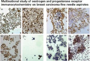
«Multinational study of oestrogen and progesterone receptor immunocytochemistry on breast carcinoma fine needle aspirates»
To collect data on the variability of immunocytochemical (ICC) procedures used to detect oestrogen/progesterone receptors (ER/PR) on cytological material; to test the reproducibility of results; and to identify the crucial points in the ICC procedures that affect the result. Methods:? Ten laboratories from eight countries participated in a two-part study. In the first part, one of the participants (the coordinator) prepared and distributed cytospins from a fine needle aspirate of a primary breast carcinoma. Laboratories performed ICC staining for ER/PR according to their own methods on the test slides and in-house positive controls. Slides were returned to the coordinator together with information on the preparation of positive control slides and the ICC methodology used. In the second part, obligatory methods of fixation and antigen retrieval were specified. Evaluation of results included grading the number of positive cells, staining intensity, background staining, cytoplasmic staining, sample condition and cellularity. Participants evaluated their own results, which were subsequently evaluated by the coordinator. Results:? There was great variability in the preparation of slides for in-house controls and ICC methodology. The outcome of ICC staining of in-house control slides was excellent in two laboratories, adequate in three, sub-optimal in four and inadequate in one. Only six obtained a positive reaction on the test slides and not all were of a high quality. Results of the second run were greatly improved in terms of cellularity of in-house positive control slides, and scores for the percentage of stained cells and staining intensity of control and test slides. Cytospins and monolayer (ThinPrep(®) ) preparations were superior to direct smears; methods of fixation and antigen retrieval were the key points in the staining process. Conclusions:? Our experience points to the need for guidelines for hormonal receptor determination and external quality control on cytological material, in order for cytological methods to be used in routine clinical practice with a suitable degree of confidence.
Journal:
Cytopathology. 2013 Feb;24(1):7-20. doi: 10.1111/cyt.12024. Epub 2012 Oct 22
Link:
http://onlinelibrary.wiley.com/doi/10.1111/cyt.12024/abstract;jsessionid...